What Is Abnormal Myocardial Perfusion
In older adults who are less active and do not exercise the heart muscle atrophy can result. Disuse or deconditioning can lead to abnormal changes in the myocardium of the older adult.
Myocardial Perfusion Imaging With Pet
To visualize blood flow patterns to the heart walls called a myocardial perfusion scan.

What is abnormal myocardial perfusion. Cardiac nuclear medicine imaging is also performed. Abnormal values of the transient ischemic dilation TID ratio are associated with severe and extensive coronary artery disease CAD. As a result under sudden emotional or physical stress the left ventricle is less able to respond to the increased demands on the myocardial muscle.
The objective of this study was to determine the relationship between TID determined from stress and rest ventricular volumes during regadenoson gated single-photon emission computed tomography myocardial perfusion. To evaluate the presence and extent of suspected or known coronary artery disease. To determine the extent of injury to the heart following a heart attack or myocardial infarction.

Solid State Detector Spect Myocardial Perfusion Imaging Journal Of Nuclear Medicine

Exercise Tl Tc Protocol Abnormal Myocardial Perfusion Tl 201 Rest Download Scientific Diagram
Understanding Your Nuclear Medicine Stress Test Digirad
2
Simulation Of Low Dose Imaging Case Example Of Abnormal Perfusion Download Scientific Diagram

Exercise Tl Tc Protocol Abnormal Myocardial Perfusion Tl 201 Rest Download Scientific Diagram
Myocardial Perfusion Spect Background Indications Contraindications

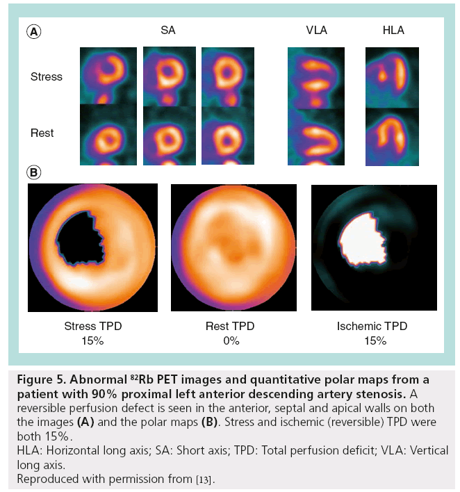
Short Axis Slices Of An Abnormal Perfusion Myocardial Study Download Scientific Diagram

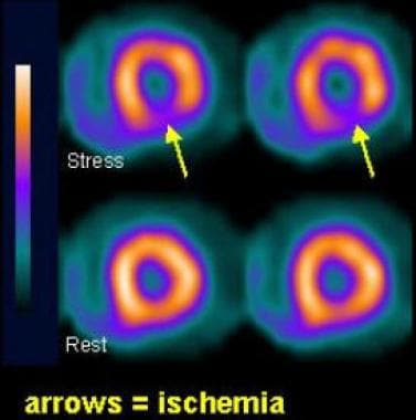
Spect Mpi A Myocardial Perfusion Study Showing A Pattern Of Reversible Download Scientific Diagram
Belum ada Komentar untuk "What Is Abnormal Myocardial Perfusion"
Posting Komentar